An open label, randomized two-way crossover scintigraphic study to investigate lung deposition of radiolabelled alginate oligosaccharide delivered as a dry powder and as a nebulized solution in cystic fibrosis patients

INTRODUCTION

Cystic fibrosis (CF) is a recessive genetic disease caused by mutations in CFTR leading to impaired
chloride and bicarbonate ion transport. This defect leads to accumulation of dense, intractable mucus
and impaired mucociliary clearance in the lungs, in turn causing inflammation, lung infections and tissue
obstruction.
OligoG CF-5/20 is a low molecular weight alginate oligosaccharide (Fig. 1) derived from brown algae,
comprised mainly of guluronate monomers. It has an inherent ability to bind divalent cations and has
been shown to disrupt bacterial biofilms in vitro and in animal models. OligoG can increase microbial
susceptibility towards antibiotics and antifungals in vitro [1-4]. It has also been shown to reduce mucus
viscosity in ex vivo CF sputum [5], and normalize the rheology of stagnant mucus in an ileal explant
model from CftrΔ508 mutant mice [6].
OligoG is currently in clinical development for cystic fibrosis, and has demonstrated excellent safety and
tolerability in healthy volunteers and CF patients.

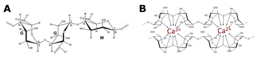
Figure 1 A: Structural composition of OligoG, showing -L-guluronate (G) and -D-manuronate
(M). At least 85% of the monomers in OligoG are G residues. B: OligoG chelating calcium

METHODS

Study design

The study was an open label two-way randomised crossover study in 10 cystic fibrosis patients. The
subjects received a single dose of OligoG CF-5/20 DPI 96 mg OligoG delivered by three capsules via the
Miat Monodose Dry Powder Inhaler, and a single dose of 1.5 mL (90 mg) aerosolised OligoG CF-5/20 6%
solution delivered via the Sidestream Plus nebuliser, separated by a 2-14 day washout period. Each
treatment was radiolabelled with 10 MBq of 99mTc in total. Technegas was drawn over a bed of DPI
using a vacuum pump, allowing the Tc to adhere to the DPI without affecting its aerodynamic properties.
Imaging
Sequential anterior and posterior images of the thorax/abdomen and lateral images of the head/neck
were acquired. Additionally, images of the device hardware were acquired pre- and post-dose, using a
Siemens E-Cam gamma camera with a 53.3 cm field of view and fitted with a low energy high-resolution
collimator.
Analysis
Image analysis was performed using the WebLink software.
Lung and extra-pulmonary deposition of radiolabel including retention in the equipment were
characterised. The effect of formulation (DPI vs solution) on the deposition parameters was assessed
using paired t-tests.

AIMS AND OBJECTIVES

Primary objective
To determine, using scintigraphic methods, the lung deposition of OligoG when administered to cystic
fibrosis patients either as a nebulised solution or as a dry powder for inhalation (DPI).
Secondary objectives
• To determine the radiolabel distribution pattern of the two formulations in the diseased lung, including
calculating the ratio of radiolabel in the central airways compared to the peripheral region (C/P index).
• To characterise the extra-pulmonary deposition (i.e. oropharyngeal and stomach) of radiolabel
including retention in the nebuliser or dry powder inhaler reservoir and deposition on the exhalation
filter.

RESULTS

* Single dose DPI inhalation was well tolerated
* Increased lung deposition of DPI as compared to the nebulised solution
(Fig.3): Mean total radioactive counts 18,766±5,798 vs 3,982±1,277
* Using the nebuliser fill volume correction factor of 1.75, the lung
deposition fraction of the 6% solution was calculated to be 17.3%, in
accordance with the 17.4% previously demonstrated [7]
* The DPI fraction deposited in the lung was 40.0±12.4% (n=10)
* Approximately 50±17% and 75±4% of the original dose remained in
the inhalers and nebuliser respectively
* Oropharyngeal deposition (Fig. 4) was significantly higher (p=0.009)
with the nebulised solution (10.1% vs 1.6%)
* The % dose in the mouth washings, that deposited in the stomach and
the calculated C/P index were not significantly different between the
two formulations

Figure 3 Deposition of OligoG in the
lungs of a CF patient

Figure 4 Deposition of OligoG in the
oropharyngeal region of a CF patient

Separate nebuliser experiment

BACKGROUND
To avoid impaired image quality due to long nebulisation time, the dose was limited to 90 mg / 1.5
mL, as compared to 270 mg /4.5 mL in the previous phase 2A. Also the nebulization time was
reduced to 2.5 min, from 15 min in 2A.
Due to increased waste using this reduced fill volume and time, a separate experiment was run to
determine the correction factor required for a realistic comparison of results from the two
formulations.
METHOD
The nebulisation equipment was set up according to the manufacturer’s instructions. The
compressor was switched on, freely generating nebulised solution. Simultaneously a stopwatch was
started (t=0). After each 30 s (for 1.5 mL samples)/1 min (for 4.5 mL samples) period, the
nebulizer chamber was detached from the tubing and weighed.
RESULTS
As deduced from Figure 2 and shown in Table 1, the counts deposited after administration of 1.5 mL
nebulised solution for 2.5 min should be multiplied with a correction factor of 1.75 in addition to the
3x, to correspond to amounts deposited from 4.5 mL nebulised for 15 min.

Table 1 Calculation of correction factor required to account for
increased waste w/1.5 mL and 2.5 min vs 4.5 mL and 15 min

Figure 2 Residual volume in nebuliser as a function of time

CONCLUSIONS

Significantly improved OligoG lung deposition (2.3 times) has been demonstrated by use of a newly
developed DPI as compared to the 6% nebulised solution.

REFERENCES

1. Khan S, et al. Overcoming Drug Resistance with Alginate Oligosaccharides Able To Potentiate the Action
of Selected Antibiotics. Antimicrob. Agents Chemother. 2012, 56(10):5134-41
2. Powell LC, et al. The effect of alginate oligosaccharides on the mechanical properties of Gram-negative
biofilms. Biofouling. 2013, 29(4):413-21
3. Roberts JL, et al. An in vitro study of alginate oligomer therapies on oral biofilms. Journal of Dentistry.
2013, 41(10):892-9
4. Powell LC, et al. A nanoscale characterization of the interaction of a novel alginate oligomer with the cell
surface and motility of Pseudomonas aeruginosa. Am J Respir Cell Mol Biol. 2014, 50(3):483-92
5. Pritchard et al. Effect of treatment with the sodium alginate oligomer OligoG, on the rheology of cystic
fibrosis sputum. ECFS 2013, WS15-5
6. Ermund A, et al. 2014, OligoG normalizes the CF mucus phenotype. See poster 235
7. Philips Respironics feasibility report on the Sidestream plus nebuliser and the 6% OligoG solution

Acknowledgements

A Dessen is the Chairman and AH Myrset is employed by AlgiPharma AS, and both are currently
shareholders of AlgiPharma; Howard Stevens is the Chairman and LA Hodges was an employee
of Bio-Images, a partner of AlgiPharma in the Eurostars project. Other authors were clinical
investigators whose hospital employer received financial contribution by AlgiPharma which was
strictly fee-for-service for conducting the study. Neither of these individuals have any
proprietary/commercial interest in OligoG or any equity interest in AlgiPharma.

CONTACT INFORMATION:

AlgiPharma AS, Industriveien 33, N-1337 Sandvika, Norway, Phone: +47 67545770

BDD Pharma Ltd, Glasgow Royal Infirmary, 84 Castle St., Glasgow G4 0SF, UK: +44 (0)141 552 8791; enquiries@bddpharma.com

LA Hodges1, G MacGregor2, JB Neilly3, HNE Stevens1, A Dessen4 and AH Myrset
4 1Bio-Images Research Ltd, Glasgow, UK; 2Gartnavel Hospital, Glasgow, UK;
3 Glasgow Royal Infirmary, Glasgow, UK;
4 AlgiPharma AS, Sandvika, Norway